BRIEF ANATOMY AND PHYSIOLOGY OF THE EYE
The eye is the primary organ of vision. Each one of the two
eyeballs is located in the orbit, where it takes up about one-fifth of the
orbital volume. The remaining space is taken up by the extraocular muscles,
fascia, fat, blood vessels, nerves and the lacrimal gland.
Basic Structure of the Eye and Supporting Structures
The Globe
The eye has three layers or coats, three compartments and
contains three fluids.
1. The three coats of the
eye are as follows:
1. (a) Outer fibrous
layer:
·
cornea
·
sclera
·
lamina cribrosa.
2. (b) Middle
vascular layer (“uveal tract”):
·
iris
·
ciliary body – consisting of the pars plicata and pars plana
·
choroids.
3. (c) Inner nervous
layer:
·
pigment epithelium of the retina
·
retinal photoreceptors
·
retinal neurons.
2. The three compartments
of the eye are as follows:
1. (a) Anterior
chamber – the space between the cornea and the iris diaphragm.
2. (b) Posterior
chamber – the triangular space between the iris anteriorly, the lens and zonule
posteriorly, and the ciliary body.
3. (c) Vitreous
chamber – the space behind the lens and zonule.
3. The three intraocular
fluids are as follows:
1. (a) Aqueous
humour – a watery, optically clear solution of water and electrolytes similar
to tissue fluids except that aqueous humour has a low protein content normally.
2. (b) Vitreous
humour – a transparent gel consisting of a three-dimensional
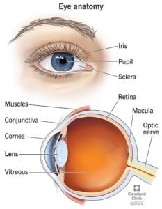
Here are descriptions of some of the main parts of the eye:
Cornea: The cornea is the clear outer part of the eye’s focusing system located
at the front of the eye.
Iris: The iris is the colored part of the eye that regulates the amount of
light entering the eye.
Lens: The lens is a clear part of the eye behind the iris that helps to focus
light, or an image, on the retina.
Macula: The macula is the small, sensitive area of the retina that gives central
vision. It is located in the center of the retina.
Optic nerve: The optic nerve is the largest sensory nerve of the eye. It
carries impulses for sight from the retina to the brain.
Pupil: The pupil is the opening at the center of the iris. The iris adjusts the
size of the pupil and controls the amount of light that can enter the eye.
Retina: The retina is the light-sensitive tissue at the back of the eye. The
retina converts light into electrical impulses that are sent to the brain
through the optic nerve.
Vitreous gel: The vitreous gel is a transparent, colorless mass that fills
the rear two-thirds of the eyeball, between the lens and the retina.
Physiology of the Eye
The primary function of the eye is to form a clear image of
objects in our environment. These images are transmitted to the brain through
the optic nerve and the posterior visual pathways. The various tissues of the
eye and its adnexa are thus designed to facilitate this function.
The Eyelids
Functions include: (1) protection of the eye from mechanical
trauma, extremes of temperature and bright light, and (2) maintenance of the
normal precorneal tear film, which is important for maintenance of corneal
health and clarity.
The Tear Film
The tear film consists of three layers: the mucoid, aqueous and
oily layers.
The mucoid layer lies adjacent to the corneal epithelium. It
improves the wetting properties of the tears. It is produced by the goblet
cells in the conjunctival epithelium.
The watery (aqueous) layer is produced by the main lacrimal
gland in the supertemporal part of the orbit and accessory lacrimal glands
found in the conjunctival stroma. This aqueous layer contains electrolytes,
proteins, lysozyme, immunoglobulins, glucose and dissolved oxygen (from the
atmosphere).
The oily layer (superficial layer of the tear film) is produced
by the meibomian glands (modified sebaceous glands) of the eyelid margins. This
oily layer helps maintain the vertical column of tears between the upper and
lower lids and prevents excessive evaporation.
The Cornea
The primary function of the cornea is refraction. In order to
perform this function, the cornea requires the following:
• transparency
• smooth and regular surface
• spherical curvature of proper refractive power
• appropriate index of refraction.
The Aqueous Humour
The aqueous humour is an optically clear solution of
electrolytes (in water) that fills the space between the cornea and the lens.
Normal volume is 0.3 ml. Its function is to nourish the lens and cornea.
The Vitreous Body
The vitreous consists of a three-dimensional network of collagen
fibres
with the interspaces filled with polymerised hyaluronic acid molecules,
which are capable of holding large quantities of water.
The Lens
The lens, like the cornea, is transparent. It is avascular and
depends on the aqueous for nourishment. It has a thick elastic capsule, which
prevents molecules (e.g., proteins) moving into or out of it.
The lens continues to grow throughout life, new lens fibres
being produced from the outside and moving inwards towards the nucleus with
age.
The lens is comprised of 65% water and 35% protein. The water
content of the lens decreases with age and the lens becomes less pliable.
The lens is suspended from the ciliary body by the zonule, which
arises from the ciliary body and inserts into the lens capsule near the
equator.
The Ciliary Body
The ciliary muscle (within the ciliary body) is a mass of smooth
muscle, which runs circumferentially inside the globe and is attached to the
scleral spur anteriorly. It consists of two main parts:
1. Longitudinal (meridional) fibres – form the outer layers and
arise from the scleral spur and insert into the choroid. Contrac- tion of this
part of the muscle exerts trac- tion on the trabecular meshwork and also the
choroid and retina.
2. Circular fibres – form the inner part and run
circumferentially. Contraction moves the ciliary processing inwards towards the
center of the pupil leading to relaxation of the zonules.
Accommodation
Accommodation is the process whereby relaxation of zonular
fibres allows the lens to become more globular, thereby increasing its
refractive power. When the ciliary muscles relax, the zonular fibres become
taut and flatten the lens, reducing its refractive power. This is associated
with constriction of the pupil and increased depth of focus.
The Retina
This is the “photographic film” of the eye that converts light
into electrical energy (transduction) for transmission to the brain. It
consists of two main parts:
1. The neuroretina – all
layers of the retina that are derived from the inner layer of the embryological
optic cup.
2. The RPE – derived from
the outer layer of the optic cup. It is comprised of a single layer of cells,
which are fixed to Bruch’s membrane. Bruch’s membrane separates the outer
retina from the choroid.


